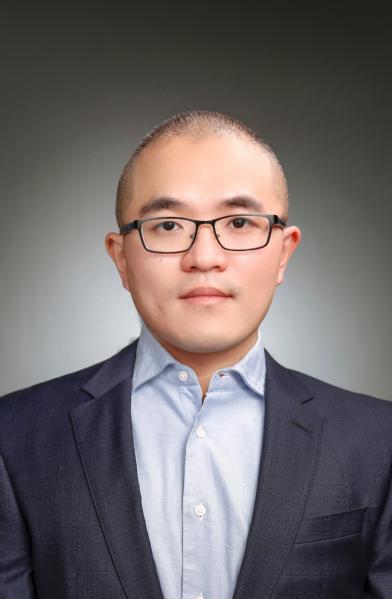
Dr. Wuwei Ren received his bachelor, master, and PhD degrees from Zhejiang University China (2010), KTH Sweden (2012), and ETH Zurich Switzerland (2018), respectively. Dr. Ren received his postdoctoral training at University of Zurich, Switzerland. He was also a visiting scholar at University College London, UK. Currently, Dr. Ren serves as a tenure-track assistant professor at ShanghaiTech University. He was awarded STINT scholarship by the Swedish government, the Swiss Innovation and Technology Agency BRIDGE Fellowship, and SNF Project Fund. In 2020, he was awarded the ‘Shanghai Youth Eastern Scholar’ Title. His work has been published in top journals such as IEEE TBME, Light, OE, BOE, JBP, Inv Prob & Img, Neurophotonics, etc. He has been actively organizing the European Molecular Imaging Meetings (EMIMs) and serving as a sub-category chair in EMIM2020 and a category chair in EMIM2021. His optical imaging prototype has won several European entrepreneurial awards including VentureKick award and displayed in Swiss Innovation Pavilion, Hannover Messe 2019. His work in system and software development has led to a PCT patent. Now he is the head of The Molecular Imaging and Biophotonics Laboratory with the aim of pioneering next-generation medical imaging tools for early diagnostics, drug development, and image-guided surgery.
Education
2012-2018 PhD in Biomedical Engineering, ETH Zurich, Switzerland
2010-2012 M. S. in Medical imaging, Royal Institute of Technology KTH, Sweden
2006-2010 B. S. in Biomedical Engineering, Zhejiang University, China
Experience
2020.9-now Assistant Professor, ShanghaiTech University, China
2020.1-2020.9 Postdoc Researcher, ETH Zurich, Switzerland
2018.8-2020.1 BRIDGE Fellow, University Hospital Zurich, Switzerland
2016.7-2016.8 Visiting Scholar, University College London, UK
Research Interests
Optical imaging (Fluorescence imaging, Optoacoustic imaging, Diffuse optical imaging, etc.)
Optical simulation, Image reconstruction & Image processing
Multimodal imaging, specialized in MRI-optics hybrid systems
Image-guided surgery & Preclinical imaging
Professional Societies
Category Chair, European Molecular Imaging Meeting (EMIM), 2021, 2020
Member of ESMI, OSA, and SPIE
President & co-founder, Swiss-Sino Medical Technology Association, 2016-2020
Honors & Awards
Shanghai Oriental Professorship, Shanghai, 2020-2023
BRIDGE fellowship, Swiss National Science Foundation, 2018-2020
Biophotonics congress travel award, OSA, 2019
STINT scholarship for academic excellence, Swedish government, 2010-2012
Outstanding student leadership award, Zhejiang University, 2010
-
Name:Liang ChenPosition:EngineerDuration:Email:
-
Name:Linlin LiPosition:PhD studentDuration:Email:
-
Name:Yexing HuPosition:Master studentDuration:Email:
-
Name:Xinyi ZhuPosition:Master studentDuration:Email:
-
Name:Chen DuanPosition:Master studentDuration:Email:
-
Name:Jianru ZhangPosition:Master studentDuration:Email:
-
Name:Yanan WuPosition:Master studentDuration:Email:
-
Name:Shihan ZhaoPosition:Master studentDuration:Email:
-
Name:Kaiqi KuangPosition:Master studentDuration:Email:
-
Name:Liangtao GuPosition:Master studentDuration:Email:
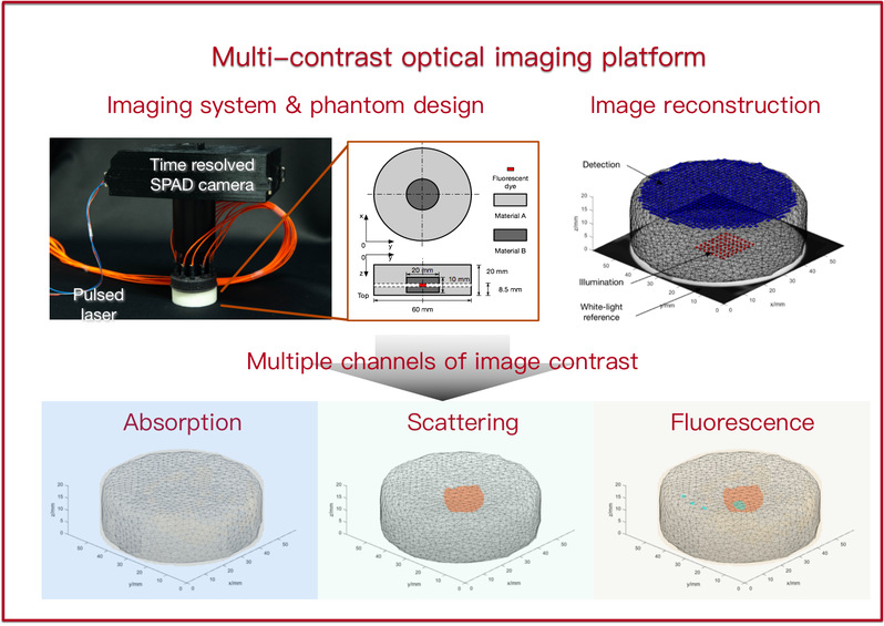
Direction 1: Multi-contrast optical imaging platform
Description: We are aiming at developing a novel optical imaging platform capable of visualizing multiple contrast parameters in biological tissues. The platform integrates the state-of-the-art hardware components such as wavelength-tunable pulse laser and time-of-flight cameras, as well as novel reconstruction algorithms and accurate calibration methods. Main application of such a platform includes small animal imaging and image-guided surgery.
Reference:
1. W. Ren, J. Jiang, A. D. Costanzo Mata, A. Kalyanov, J. Ripoll, S. Lindner, E. Charbon, C. Zhang, M. Rudin, and M. Wolf, Multimodal imaging combining time-domain near-infrared optical tomography and continuous-wave fluorescence molecular tomography, Opt Express 28, 9860-9874 (2020).
2. W. Ren, H. Isler, M. Wolf, J. Ripoll, and M. Rudin, Smart Toolkit for Fluorescence Tomography: Simulation, Reconstruction, and Validation, IEEE Trans Biomed Eng 67, 16-26 (2020).
3. W. Ren, L. Li, J. Zhang, M. Vaas, J. Klohs, J. Ripoll, M. Wolf, R. Ni, and M. Rudin, Non-invasive visualization of amyloid-beta deposits in Alzheimer amyloidosis mice using magnetic resonance imaging and fluorescence molecular tomography, Biomed Opt Express 13, 3809-3822 (2022).
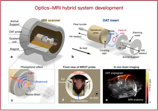
Direction 2: Optics-MRI hybrid system
Description: Multimodal imaging has emerged as a powerful method for biomedical research and clinical usage, as more comprehensive physiological information can be achieved compared with a standalone modality. In our lab, we combine optics and MRI due to several reasons. First, optical imaging, fluorescence imaging in particular, features high sensitivity and specificity which is complementary to MRI. Second, MRI can provides as an accurate anatomical reference that is useful for localizing the fluorescence signal. Third, both MRI and optical imaging can avoid ionizing radiation. Our team has previously developed the world’s first fluorescence tomography-MRI system and optoacoustic tomography-MRI system.
Reference:
1. W. Ren, X. L. Dean-Ben, M. A. Augath, and D. Razansky, Development of concurrent magnetic resonance imaging and volumetric optoacoustic tomography: A phantom feasibility study, J Biophotonics 14, e202000293 (2021).
2. Y. Hu, B. Lafci, A. Luzgin, H. Wang, J. Klohs, X. L. Dean-Ben, R. Ni, D. Razansky, and W. Ren, Deep learning facilitates fully automated brain image registration of optoacoustic tomography and magnetic resonance imaging, Biomed Opt Express 13, 4817-4833 (2022).
3. W. Ren, B. Ji, Y. Guan, L. Cao, and R. Ni, Recent Technical Advances in Accelerating the Clinical Translation of Small Animal Brain Imaging: Hybrid Imaging, Deep Learning, and Transcriptomics, Front Med (Lausanne) 9, 771982 (2022).
Direction 3: AI-empowered image reconstruction
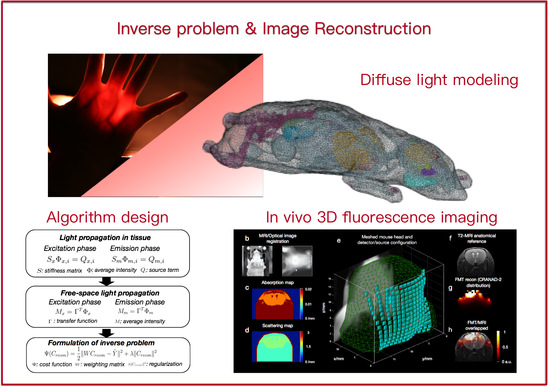
Description: The conception of 3D reconstruction is commonly used in optical microscopy, e.g., in confocal microscopy and two-photon microscopy. However, visualizing objects deeply seated in tissue (> 1 mm) becomes challenging due to severe light scattering in tissue. Such a macroscopic reconstruction in scattering media is well known as a highly ill-posed inverse problem. We are seeking for fast, robust, 3D, high-resolution image reconstruction algorithms based on advanced computational techniques and novel machine learning methods.
Reference:
1. Y. Liu, W. Ren, and H. Ammari, Robust reconstruction of fluorescence molecular tomography with an optimized illumination pattern, Inverse Problems & Imaging 14, 535-568 (2020).
2. W. Ren, H. Isler, M. Wolf, J. Ripoll, and M. Rudin, Smart Toolkit for Fluorescence Tomography: Simulation, Reconstruction, and Validation, IEEE Trans Biomed Eng 67, 16-26 (2020).
Digital Signal Processing, ShanghaiTech-EE152, 2021-2025 Spring
Digital Image Processing, ShanghaiTech-CS270, 2021-2023 Fall
Multi-modal Brain-Computer Interface, ShanghaiTech-SI361, 2021-2024 Spring
New! Biomedical photonics and imaging, ShanghaiTech-EE280, 2024 Fall
著作Book Chapter:
Ren W*, Ni R* (2024) Chapter 16: Noninvasive Visualization of Amyloid-Beta Deposits in Alzheimer’s Amyloidosis Mice via Fluorescence Molecular Tomography Using Contrast Agent. Book: Biomarkers for Alzheimer’s Disease Drug Development 2785: 271-285, Springer
期刊论文Journal papers:
Shen S#, Li L#, Gao S, Wang Y, Gu L, Li S, Zhu X, Jiang J*, Yu J*, Ren W* (2024) High Resolution Diffuse Optical Tomography via Neural Fields, IEEE Transactions on Computational Imaging
Landman MS*, Jiang J*, Zhang J, Ren W (2024) Augmented Flexible Krylov Subspace methods with applications to Bayesian inverse problems. Linear Algebra and its Applications
Wu Y, Hu Y, Li L, Zhu X, Gu L, Jiang J, Ren W* (2024) Simultaneous reconstruction of 3D fluores- cence distribution and object surface using structured light illumination and dual-camera detection. Optics Express 32(7)
Wang J, Pan B, Wang Z, Zhang J, Zhou Z, Yao L, Wu Y, Ren W, Wang J, Ji H, Yu J*, Chen B* (2024) Single-pixel p-graded-n junction spectrometers. Nat Commun 15: 1773
Wu Y, Li F, Wu Y, Wang H, Gu L, Zhang J, Qi Y, Meng L, Kong N, Chai Y, Hu Q, Xing Z, Ren W*,Li F*, Zhu X* (2024) Lanthanide luminescence nanothermometer with working wavelength beyond 1500 nm for cerebrovascular temperature imaging in vivo. Nat Commun 15: 2341
Zhang J, Wang Z, Cao T, Cao G*, Ren W*, Jiang J* (2024) Robust residual-guided iterative reconstruction for sparse-view CT in small animal imaging. Physics in Medicine & Biology
Zhu X, Gu L, Li R, Chen L, Chen, J, Zhou N*, Ren W* (2023), MiniMounter: a low‐cost miniaturized microscopy development toolkit for image quality control and enhancement. Journal of Biophotonics, e202300214
Gao S, Zhang J, Hu Y, Wu Y, Li L, Hu Q, Lou X, Zhu X, Jiang J*, Ren W* (2023), Multifunctional Optical Tomography System with High-fidelity Surface Extraction based on a Single Programmable Scanner and Unified Pinhole Modeling, IEEE Transactions on Biomedical Engineering, 1-13
Ren W, Dean-Ben Xl, Skachokova Z, Augath M, Ni R, Chen Z, Razansky D* (2023) Monitoring mouse brain perfusion with hybrid magnetic resonance optoacoustic tomography. Biomed Opt Express 14: 1192-1204
Ren W*, Li L, Zhang J, Vaas M, Klohs J, Ripoll J, Wolf M, Ni R, Rudin M (2022) Non-invasive visualization of amyloid-beta deposits in Alzheimer amyloidosis mice using magnetic resonance imaging and fluorescence molecular tomography. Biomed Opt Express 13: 3809-3822
Ren W*, Ji B, Guan Y, Cao L, Ni R* (2022) Recent Technical Advances in Accelerating the Clinical Translation of Small Animal Brain Imaging: Hybrid Imaging, Deep Learning, and Transcriptomics. Front Med (Lausanne) 9: 771982
Hu Y, Lafci B, Luzgin A, Wang H, Klohs J, Dean-Ben XL, Ni R, Razansky D*, Ren W*(2022) Deep learning facilitates fully automated brain image registration of optoacoustic tomography and magnetic resonance imaging. Biomed Opt Express 13: 4817-4833
Chen Z, Gezginer I, Augath MA, Ren W, Liu YH, Ni R, Dean-Ben XL, Razansky D* (2022) Hybrid magnetic resonance and optoacoustic tomography (MROT) for preclinical neuroimaging. Light Sci Appl 11: 332
Zhou R, Ji B, Kong Y, Qin L, Ren W, Guan Y, Ni R* (2021) PET Imaging of Neuroinflammation in Alzheimer's Disease. Front Immunol 12: 739130
Ren W, Dean-Ben XL, Augath MA, Razansky D* (2021) Development of concurrent magnetic resonance imaging and volumetric optoacoustic tomography: A phantom feasibility study. J Biophotonics 14: e202000293
Ren W, Cui S, Alini M, Grad S, Zhou Q, Li Z, Razansky D* (2021) Noninvasive multimodal fluorescence and magnetic resonance imaging of whole-organ intervertebral discs. Biomed Opt Express 12: 3214-3227
Ren W*, Jiang J, Costanzo Mata AD, Kalyanov A, Ripoll J, Lindner S, Charbon E, Zhang C, Rudin M, Wolf M (2020) Multimodal imaging combining time-domain near-infrared optical tomography and continuous-wave fluorescence molecular tomography. Opt Express 28: 9860-9874
Ren W, Isler H, Wolf M, Ripoll J, Rudin M* (2020) Smart Toolkit for Fluorescence Tomography: Simulation, Reconstruction, and Validation. IEEE Trans Biomed Eng 67: 16-26
Liu Y, Ren W*, Ammari H (2020) Robust reconstruction of fluorescence molecular tomography with an optimized illumination pattern. Inverse Problems & Imaging 14: 535-568
Ren W, Skulason H, Schlegel F, Rudin M, Klohs J, Ni R* (2019) Automated registration of magnetic resonance imaging and optoacoustic tomography data for experimental studies. Neurophotonics 6: 025001
Ni R, Vaas M, Ren W, Klohs J* (2018) Noninvasive detection of acute cerebral hypoxia and subsequent matrix-metalloproteinase activity in a mouse model of cerebral ischemia using multispectral-optoacoustic-tomography. Neurophotonics 5: 015005
Ren W, Elmer A, Buehlmann D, Augath M-A, Vats D, Ripoll J, Rudin M* (2016) Dynamic Measurement of Tumor Vascular Permeability and Perfusion using a Hybrid System for Simultaneous Magnetic Resonance and Fluorescence Imaging. Mol Imaging Biol 18: 191-200
会议论文Proceeding papers:
Hu Y, Lafci B, Luzgin A, Wang H, Klohs J, Dean-Ben XL, Ni R, Razansky D, Ren W* (2022) Automatic image registration of optoacoustic tomography and magnetic resonance imaging based on deep learning. Proc. SPIE, First Conference on Biomedical Photonics and Cross-Fusion, 1246103, doi: doi.org/10.1117/12.2655214
Ren W, Dean-Ben XL, Augath M, Razansky D* (2021) Feasibility study on concurrent optoacoustic tomography and magnetic resonance imaging. Proc. SPIE, doi: (oral & Best Paper Nomination in SPIE Photonics West, ranking top 5%)
Ren W*, Jiang J, Di Costanzo MA, Kalyanov A, Ripoll J, Lindner S, Charbon S, Zhang C, Rudin M, Wolf M (2020) Time-domain Near Infrared Optical Tomography Guided Fluorescence Molecular Tomography, Biophotonics Congress: Biomedical Optic. OSA
Jiang J, Ren W, Isler H, Kalyanov A, Lindner S, Mata Aldo D. C, Rudin M, Wolf M* (2020), Validation and Comparison of Monte Carlo and Finite Element Method in Forward Modeling for Near Infrared Optical Tomography. In Oxygen Transport to Tissue XLI (pp. 307-313). Springer, Charm
Polatoglu M N, Liu Y, Ni R, Ripoll J, Rudin M, Wolf M, Ren W* (2019), Simulation of fluorescence molecular tomography using a registered digital mouse atlas, Proc. SPIE, doi: /10.1117/12.2525366
Ren W, Skulason H, Schlegel F, Rudin M, Klohs J, Ni R* (2019), Automated registration for optoacoustic tomography and MRI, In Optical Molecular Probes, Imaging and Drug Delivery. OSA
Ni R, Vaas M, Ren W, Klohs J* (2018), Non-invasive detection of matrix-metalloproteinase activity in a mouse model of cerebral ischemia using multispectral optoacoustic tomography, Proc. SPIE, doi: 10.1117/12.2286313
Ren W, Elmer A, Augath M, Rudin M* (2016), FEM-based Simulation of a Fluorescence Tomography Experiment using Anatomical MR Images, Proc. SPIE, doi:10.1117/12.2216454
Ren W, Valastyán I, Colarieti-Tosti M* (2012), Stationary SPECT with multi-layer Multiple-Pinhole-Arrays, IEEE Nuclear Science Symposium and Medical Imaging Conference (NSS/MIC), doi: 10.1109/NSSMIC.2012.6551593
Valastyán I*, Colarieti-Tostia M, Ren W, Turco, A, Kereka A (2012), Monte Carlo simulation of a dental Positron Emission Tomograph and image reconstruction of scatter and true coincidence events, IEEE Nuclear Science Symposium and Medical Imaging Conference (NSS/MIC), doi: 10.1109/NSSMIC.2012.6551795
专利Patent:
Ren W, Rudin M, Method for selecting an illumination pattern for a fluorescence molecular tomography measurement. PCT/EP2020/051779
Ren W, Zhu X, Chen L. Microscopic Device for small animal imaging (动物显微成像设备). CN118112769A
Ren W, Zhu X, Gu L. Imaging device combining MRI and optics (结合核磁共振和光学的小动物成像系统). CN118161144A
Ren W, Wu Y, Hu Y. Simultaneous data acquisition system for 3D fluorescence visualization and surface extraction (同时提取内部荧光分子分布和表面三维结构的成像系统). CN116839500A
Ren W, Zhu X, Chen L, Gu L. Calibration tool for miniaturized microscopy (适用于微型化显微成像的辅助调节系统). CN116661122A
Ren W, Wu Y, Chen L. Duo-view surface extraction and optical tomography using a single camera (单相机提取双视角表面信息的光学断层扫描系统). CN116576796A
Ren W, Gao S, Wu Y. Super-continuous wave source non-contact optical tomography (连续波光源非接触式的光学断层扫描系统及扫描方法). CN115191947A


 沪公网安备 31011502006855号