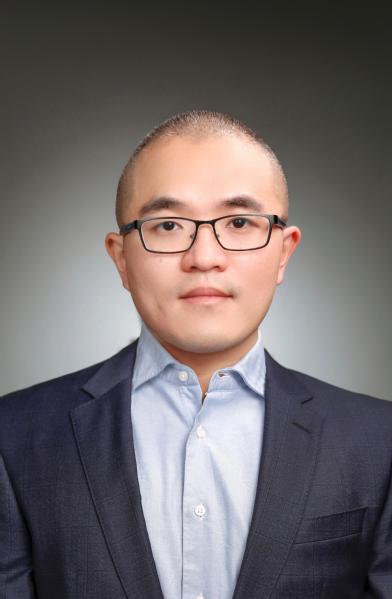
任无畏,上海科技大学信息学院助理教授/研究员/博导,研究兴趣包括光学成像、图像重建、多模态融合、以及荧光手术导航和小动物成像等应用场景。曾就读于浙江大学(2010)、瑞典皇家理工学院(2012)、瑞士苏黎世联邦理工学院(2018)并分别获得学士、硕士、博士学位。2016年赴英国伦敦大学学院医学图像计算中心访学,后在瑞士苏黎世大学医学院及药学院完成博士后工作。主要学术成果发表在Nature Comm、TBME、Light、OE、BOE、JBP、PMB等知名期刊。任博士自主研发的光学成像原型机曾斩获VentureKick等欧洲知名创业奖项,并作为瑞士创业代表参展2019年度德国汉诺威工业展。近五年来申报中国专利8项,国际专利(PCT)1项。获瑞典政府STINT全额奖学金,瑞士创新科技署BRIDGE成果转化基金(首位华人得奖者),瑞士国家科学基金会基础项目基金,国自然青年项目基金,入选上海市“青年东方学者”人才计划和上海市领军人才计划。目前担任OPTICA,IEEE等多个协会旗下期刊审稿人,以及欧洲分子成像年会分会主席。
教育经历
2012-2018 博士,生物医学工程专业,瑞士苏黎世联邦理工学院
2010-2012 硕士,生物医学工程专业,瑞典皇家理工学院
2006-2010 学士,生物医学工程专业,浙江大学
工作经历
2020.9-now 助理教授,上海科技大学
2020.1-2020.9 博士后,生物医学工程专业,瑞士苏黎世联邦理工学院
2018.8-2020.1 博士后,瑞士苏黎世大学医院
2016.7-2016.8 访问学者,英国伦敦大学学院
学生培养
本实验室欢迎对成像科学有浓厚兴趣的同学加入!专业背景包括计算机、电子、应用数学和光学工程等。毕业学生去向包括海外深造(新加坡南洋理工大学、英国伯明翰大学全额奖学金博士)、国内知名医疗器械公司(如联影)。
-
姓名:陈亮身份:工程师年级:邮箱:研究方向:
-
姓名:李林霖身份:博士研究生年级:邮箱:研究方向:
-
姓名:胡叶兴身份:硕士研究生年级:邮箱:研究方向:
-
姓名:邾馨怡身份:硕士研究生年级:邮箱:研究方向:
-
姓名:段晨身份:硕士研究生年级:邮箱:研究方向:
-
姓名:张柬如身份:硕士研究生年级:邮箱:研究方向:
-
姓名:吴亚男身份:硕士研究生年级:邮箱:研究方向:
-
姓名:赵世涵身份:硕士研究生年级:邮箱:研究方向:
-
姓名:匡恺琦身份:硕士研究生年级:邮箱:研究方向:
-
姓名:顾梁韬身份:硕士研究生年级:邮箱:研究方向:
方向一:多对比度光学成像系统
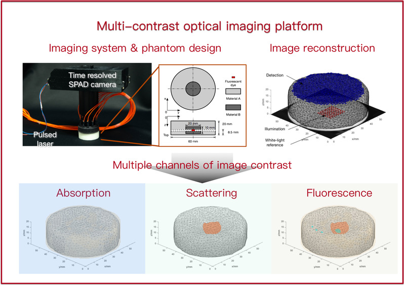
我们的目标是开发一种能够可视化生物组织中多个对比度参数的新型光学成像平台。 该平台集成了波长可调脉冲激光器和飞行时间相机等最先进的硬件组件,以及新颖的重建算法和精确的校准方法。 这种平台的主要应用包括小动物成像和图像引导手术。
参考文献:
1. W. Ren, J. Jiang, A. D. Costanzo Mata, A. Kalyanov, J. Ripoll, S. Lindner, E. Charbon, C. Zhang, M. Rudin, and M. Wolf, Multimodal imaging combining time-domain near-infrared optical tomography and continuous-wave fluorescence molecular tomography, Opt Express 28, 9860-9874 (2020).
2. W. Ren, H. Isler, M. Wolf, J. Ripoll, and M. Rudin, Smart Toolkit for Fluorescence Tomography: Simulation, Reconstruction, and Validation, IEEE Trans Biomed Eng 67, 16-26 (2020).
3. W. Ren, L. Li, J. Zhang, M. Vaas, J. Klohs, J. Ripoll, M. Wolf, R. Ni, and M. Rudin, Non-invasive visualization of amyloid-beta deposits in Alzheimer amyloidosis mice using magnetic resonance imaging and fluorescence molecular tomography, Biomed Opt Express 13, 3809-3822 (2022).

方向二:光学-MRI混合系统
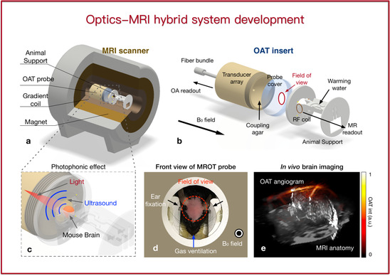
多模态成像已成为生物医学研究和临床使用的一种强大方法。与单一模态相比,可以获得更全面的生理信息。我们将光学和 MRI 相结合有多方面的优点。 首先,光学成像,特别是荧光成像,具有高灵敏度和特异性,与MRI高度互补。 其次,MRI 可以提供准确的解剖学参考,有助于定位荧光信号。 第三,MRI和光学成像都可以避免电离辐射。 我们的团队此前开发了世界上第一台荧光断层扫描-MRI系统和光声断层扫描-MRI系统。
参考文献:
1. W. Ren, X. L. Dean-Ben, M. A. Augath, and D. Razansky, Development of concurrent magnetic resonance imaging and volumetric optoacoustic tomography: A phantom feasibility study, J Biophotonics 14, e202000293 (2021).
2. Y. Hu, B. Lafci, A. Luzgin, H. Wang, J. Klohs, X. L. Dean-Ben, R. Ni, D. Razansky, and W. Ren, Deep learning facilitates fully automated brain image registration of optoacoustic tomography and magnetic resonance imaging, Biomed Opt Express 13, 4817-4833 (2022).
3. W. Ren, B. Ji, Y. Guan, L. Cao, and R. Ni, Recent Technical Advances in Accelerating the Clinical Translation of Small Animal Brain Imaging: Hybrid Imaging, Deep Learning, and Transcriptomics, Front Med (Lausanne) 9, 771982 (2022).

方向三:AI赋能的图像重建
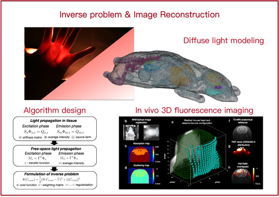
3D 重建的概念通常用于光学显微镜,例如共聚焦显微镜和双光子显微镜。 然而,由于组织中严重的光散射,可视化深入组织(> 1 毫米)的物体变得具有挑战性。 众所周知,散射介质中的这种宏观重建是高度不适定的逆问题。 我们正在寻求基于先进计算技术和新颖机器学习方法的快速、稳健、3D、高分辨率图像重建算法。
参考文献:
1. Y. Liu, W. Ren, and H. Ammari, Robust reconstruction of fluorescence molecular tomography with an optimized illumination pattern, Inverse Problems & Imaging 14, 535-568 (2020).
2. W. Ren, H. Isler, M. Wolf, J. Ripoll, and M. Rudin, Smart Toolkit for Fluorescence Tomography: Simulation, Reconstruction, and Validation, IEEE Trans Biomed Eng 67, 16-26 (2020).

数字信号处理, ShanghaiTech-EE152, 2021-2025 Spring
数字图像处理, ShanghaiTech-CS270, 2021-2023 Fall
多模态脑机接口, ShanghaiTech-SI361, 2021-2024 Spring
New! 生物医学光子学及成像, ShanghaiTech-EE280, 2024 Fall
著作Book Chapter:
Ren W*, Ni R* (2024) Chapter 16: Noninvasive Visualization of Amyloid-Beta Deposits in Alzheimer’s Amyloidosis Mice via Fluorescence Molecular Tomography Using Contrast Agent. Book: Biomarkers for Alzheimer’s Disease Drug Development 2785: 271-285, Springer
期刊论文Journal papers:
[Highlight] Shen S#, Li L#, Gao S, Wang Y, Gu L, Li S, Zhu X, Jiang J*, Yu J*, Ren W* (2024) High Resolution Diffuse Optical Tomography via Neural Fields, IEEE Transactions on Computational Imaging
Landman MS*, Jiang J*, Zhang J, Ren W (2024) Augmented Flexible Krylov Subspace methods with applications to Bayesian inverse problems. Linear Algebra and its Applications
[Highlight] Wu Y, Hu Y, Li L, Zhu X, Gu L, Jiang J, Ren W* (2024) Simultaneous reconstruction of 3D fluores- cence distribution and object surface using structured light illumination and dual-camera detection. Optics Express 32(7)
Wang J, Pan B, Wang Z, Zhang J, Zhou Z, Yao L, Wu Y, Ren W, Wang J, Ji H, Yu J*, Chen B* (2024) Single-pixel p-graded-n junction spectrometers. Nat Commun 15: 1773
[Highlight] Wu Y, Li F, Wu Y, Wang H, Gu L, Zhang J, Qi Y, Meng L, Kong N, Chai Y, Hu Q, Xing Z, Ren W*,Li F*, Zhu X* (2024) Lanthanide luminescence nanothermometer with working wavelength beyond 1500 nm for cerebrovascular temperature imaging in vivo. Nat Commun 15: 2341
Zhang J, Wang Z, Cao T, Cao G*, Ren W*, Jiang J* (2024) Robust residual-guided iterative reconstruction for sparse-view CT in small animal imaging. Physics in Medicine & Biology
Zhu X, Gu L, Li R, Chen L, Chen, J, Zhou N*, Ren W* (2023), MiniMounter: a low‐cost miniaturized microscopy development toolkit for image quality control and enhancement. Journal of Biophotonics, e202300214
[Highlight] Gao S, Zhang J, Hu Y, Wu Y, Li L, Hu Q, Lou X, Zhu X, Jiang J*, Ren W* (2023), Multifunctional Optical Tomography System with High-fidelity Surface Extraction based on a Single Programmable Scanner and Unified Pinhole Modeling, IEEE Transactions on Biomedical Engineering, 1-13
Ren W, Dean-Ben Xl, Skachokova Z, Augath M, Ni R, Chen Z, Razansky D* (2023) Monitoring mouse brain perfusion with hybrid magnetic resonance optoacoustic tomography. Biomed Opt Express 14: 1192-1204
Ren W*, Li L, Zhang J, Vaas M, Klohs J, Ripoll J, Wolf M, Ni R, Rudin M (2022) Non-invasive visualization of amyloid-beta deposits in Alzheimer amyloidosis mice using magnetic resonance imaging and fluorescence molecular tomography. Biomed Opt Express 13: 3809-3822
Ren W*, Ji B, Guan Y, Cao L, Ni R* (2022) Recent Technical Advances in Accelerating the Clinical Translation of Small Animal Brain Imaging: Hybrid Imaging, Deep Learning, and Transcriptomics. Front Med (Lausanne) 9: 771982
Hu Y, Lafci B, Luzgin A, Wang H, Klohs J, Dean-Ben XL, Ni R, Razansky D*, Ren W*(2022) Deep learning facilitates fully automated brain image registration of optoacoustic tomography and magnetic resonance imaging. Biomed Opt Express 13: 4817-4833
Chen Z, Gezginer I, Augath MA, Ren W, Liu YH, Ni R, Dean-Ben XL, Razansky D* (2022) Hybrid magnetic resonance and optoacoustic tomography (MROT) for preclinical neuroimaging. Light Sci Appl 11: 332
Zhou R, Ji B, Kong Y, Qin L, Ren W, Guan Y, Ni R* (2021) PET Imaging of Neuroinflammation in Alzheimer's Disease. Front Immunol 12: 739130
Ren W, Dean-Ben XL, Augath MA, Razansky D* (2021) Development of concurrent magnetic resonance imaging and volumetric optoacoustic tomography: A phantom feasibility study. J Biophotonics 14: e202000293
Ren W, Cui S, Alini M, Grad S, Zhou Q, Li Z, Razansky D* (2021) Noninvasive multimodal fluorescence and magnetic resonance imaging of whole-organ intervertebral discs. Biomed Opt Express 12: 3214-3227
Ren W*, Jiang J, Costanzo Mata AD, Kalyanov A, Ripoll J, Lindner S, Charbon E, Zhang C, Rudin M, Wolf M (2020) Multimodal imaging combining time-domain near-infrared optical tomography and continuous-wave fluorescence molecular tomography. Opt Express 28: 9860-9874
Ren W, Isler H, Wolf M, Ripoll J, Rudin M* (2020) Smart Toolkit for Fluorescence Tomography: Simulation, Reconstruction, and Validation. IEEE Trans Biomed Eng 67: 16-26
Liu Y, Ren W*, Ammari H (2020) Robust reconstruction of fluorescence molecular tomography with an optimized illumination pattern. Inverse Problems & Imaging 14: 535-568
Ren W, Skulason H, Schlegel F, Rudin M, Klohs J, Ni R* (2019) Automated registration of magnetic resonance imaging and optoacoustic tomography data for experimental studies. Neurophotonics 6: 025001
Ni R, Vaas M, Ren W, Klohs J* (2018) Noninvasive detection of acute cerebral hypoxia and subsequent matrix-metalloproteinase activity in a mouse model of cerebral ischemia using multispectral-optoacoustic-tomography. Neurophotonics 5: 015005
Ren W, Elmer A, Buehlmann D, Augath M-A, Vats D, Ripoll J, Rudin M* (2016) Dynamic Measurement of Tumor Vascular Permeability and Perfusion using a Hybrid System for Simultaneous Magnetic Resonance and Fluorescence Imaging. Mol Imaging Biol 18: 191-200
会议论文Proceeding papers:
Hu Y, Lafci B, Luzgin A, Wang H, Klohs J, Dean-Ben XL, Ni R, Razansky D, Ren W* (2022) Automatic image registration of optoacoustic tomography and magnetic resonance imaging based on deep learning. Proc. SPIE, First Conference on Biomedical Photonics and Cross-Fusion, 1246103, doi: doi.org/10.1117/12.2655214
Ren W, Dean-Ben XL, Augath M, Razansky D* (2021) Feasibility study on concurrent optoacoustic tomography and magnetic resonance imaging. Proc. SPIE, doi: (oral & Best Paper Nomination in SPIE Photonics West, ranking top 5%)
Ren W*, Jiang J, Di Costanzo MA, Kalyanov A, Ripoll J, Lindner S, Charbon S, Zhang C, Rudin M, Wolf M (2020) Time-domain Near Infrared Optical Tomography Guided Fluorescence Molecular Tomography, Biophotonics Congress: Biomedical Optic. OSA
Jiang J, Ren W, Isler H, Kalyanov A, Lindner S, Mata Aldo D. C, Rudin M, Wolf M* (2020), Validation and Comparison of Monte Carlo and Finite Element Method in Forward Modeling for Near Infrared Optical Tomography. In Oxygen Transport to Tissue XLI (pp. 307-313). Springer, Charm
Polatoglu M N, Liu Y, Ni R, Ripoll J, Rudin M, Wolf M, Ren W* (2019), Simulation of fluorescence molecular tomography using a registered digital mouse atlas, Proc. SPIE, doi: /10.1117/12.2525366
Ren W, Skulason H, Schlegel F, Rudin M, Klohs J, Ni R* (2019), Automated registration for optoacoustic tomography and MRI, In Optical Molecular Probes, Imaging and Drug Delivery. OSA
Ni R, Vaas M, Ren W, Klohs J* (2018), Non-invasive detection of matrix-metalloproteinase activity in a mouse model of cerebral ischemia using multispectral optoacoustic tomography, Proc. SPIE, doi: 10.1117/12.2286313
Ren W, Elmer A, Augath M, Rudin M* (2016), FEM-based Simulation of a Fluorescence Tomography Experiment using Anatomical MR Images, Proc. SPIE, doi:10.1117/12.2216454
Ren W, Valastyán I, Colarieti-Tosti M* (2012), Stationary SPECT with multi-layer Multiple-Pinhole-Arrays, IEEE Nuclear Science Symposium and Medical Imaging Conference (NSS/MIC), doi: 10.1109/NSSMIC.2012.6551593
Valastyán I*, Colarieti-Tostia M, Ren W, Turco, A, Kereka A (2012), Monte Carlo simulation of a dental Positron Emission Tomograph and image reconstruction of scatter and true coincidence events, IEEE Nuclear Science Symposium and Medical Imaging Conference (NSS/MIC), doi: 10.1109/NSSMIC.2012.6551795
专利Patent:
Ren W, Rudin M, Method for selecting an illumination pattern for a fluorescence molecular tomography measurement. PCT/EP2020/051779
Ren W, Zhu X, Chen L. Microscopic Device for small animal imaging (动物显微成像设备). CN118112769A
Ren W, Zhu X, Gu L. Imaging device combining MRI and optics (结合核磁共振和光学的小动物成像系统). CN118161144A
Ren W, Wu Y, Hu Y. Simultaneous data acquisition system for 3D fluorescence visualization and surface extraction (同时提取内部荧光分子分布和表面三维结构的成像系统). CN116839500A
Ren W, Zhu X, Chen L, Gu L. Calibration tool for miniaturized microscopy (适用于微型化显微成像的辅助调节系统). CN116661122A
Ren W, Wu Y, Chen L. Duo-view surface extraction and optical tomography using a single camera (单相机提取双视角表面信息的光学断层扫描系统). CN116576796A
Ren W, Gao S, Wu Y. Super-continuous wave source non-contact optical tomography (连续波光源非接触式的光学断层扫描系统及扫描方法). CN115191947A


 沪公网安备 31011502006855号


