
With the advantages of non-radiation, non-invasive and flexible operation, the 3D ultrasound imaging technique has been greatly refined and applied in medical imaging and diagnosis. In general, the 3D ultrasound imaging system is comprised of a conventional ultrasound probe and a location sensor, which can generate 2D transverse images and the corresponding spatial information needed for the subsequent reconstruction process of rebuilding these data into a 3D space. However, to scan a complete spine, it may generate as many as 2000 2D images so that reconstructing a 3D image using such a huge number of 2D images is time-consuming. This problem greatly limits this technique’s application in broader clinical applications.
To overcome this limitation, Assistant Professor Zheng Rui’s research group in the Smart Medical Information Research Center (SMIRC) of SIST proposed a Fast-Dot Projection (FDP) algorithm to speed up the reconstruction process. In this algorithm, the complex vector operations used in traditional reconstruction algorithms are replaced by numerical operations to simplify and accelerate the projection process, so that this algorithm can be employed universally for different reconstruction methods. The researchers validated FDP by using the datasets of scoliosis patients. Their achievement was recently published in IEEE Transactions on Ultrasonics, Ferroelectrics, and Frequency Control (IEEE TUFFC) in an article entitled “Improvement of 3D Ultrasound Spine Imaging technique using Fast Reconstruction Algorithm”. Meanwhile, this article has been selected as the Front Cover of the October issue.
The FDP algorithm has three outstanding advantages. First, it can be adapted to various reconstruction methods such as voxel-based back mapping or pixel-based forward mapping. Second, the fusion speed for 2D images with spatial information is much faster, with the fastest reconstruction time dropping to 26 seconds, only one fifth the time taken by conventional methods, making more complex reconstruction methods applicable in clinical settings which are designed to acquire higher quality images. Third, due to the much shorter computation time, real-time visualization of the reconstructed results can be achieved, thereby enabling simultaneous clinical diagnosis and assessment while scanning the patients.
Because of the real-time imaging ability, this achievement has brought the development and application of 3D ultrasound spine imaging into clinical practice. It makes possible the replacement of the current X-ray imaging method with 3D ultrasound imaging as a non-radiation diagnostic technique, laying a foundation for mass screening of scoliosis and other diseases.
Second year Ph.D. candidate Chen Hongbo is the first author of this article. Prof. Zheng is the corresponding author.
Link to the Front cover:
https://ieeexplore.ieee.org/stamp/stamp.jsp?tp=&arnumber=9548855
Article link:
https://ieeexplore.ieee.org/document/9449836
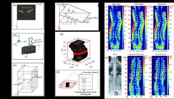
Left: the schematic diagram of reconstruction; middle: the schematic diagram of algorithms; right: the imaging results
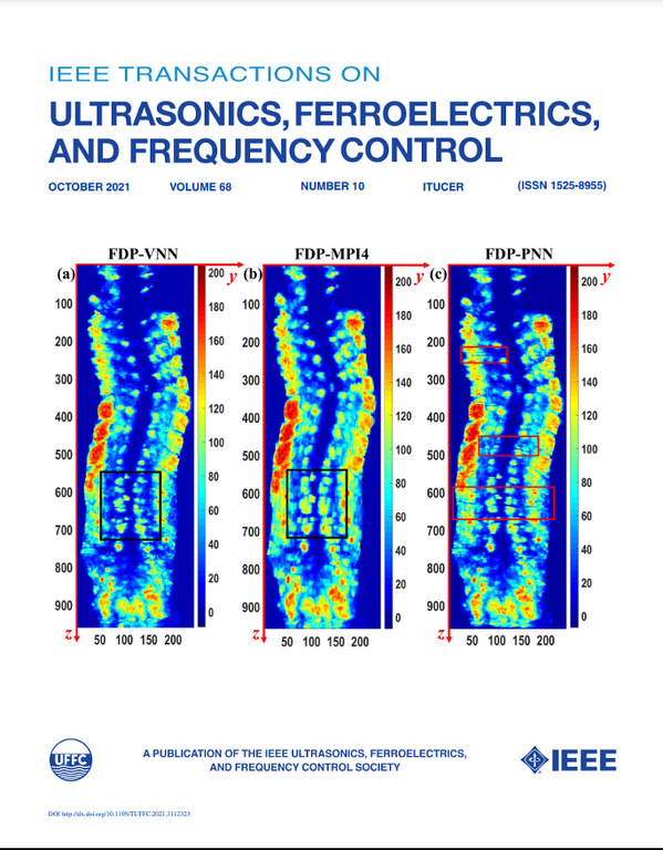
Front Cover

 沪公网安备 31011502006855号