Microbubble cavitation-based focused ultrasound therapies can non-invasively open biological barriers temporarily, such as the blood-brain barrier and blood-tumor barrier. This emerging new type of therapy is undergoing a huge number of clinical investigations for drug delivery and for removing pathological tissues without opening the human body, and has great potential for the treatment of brain diseases and cancer. Passive acoustic mapping (PAM) is a promising tool used to monitor microbubble cavitation activities during therapy, in order to guarantee the safety and effectiveness of the treatment. However, the existing PAM methods for convex arrays rely on the computationally expensive delay-and-sum (DAS) operation so that the image reconstruction speed is limited when the field-of-view is large.
To address this issue, Assistant Professor Cai Xiran’s group in the School of Information Science and Technology (SIST) proposed a fast beamforming method for PAM with convex arrays, achieving an image reconstruction speed at the millisecond level. This method is based on the projection of the helical wave spectrum (HWS) between cylindrical surfaces in the imaging area (Figure 1). The proposed method has image quality and acoustic source localization accuracy comparable with the classical DAS-based methods, as validated through simulations and in vitro experiments, while the image reconstruction speed has been remarkably improved, by more than tenfold.
Additionally, the frequency selectivity feature of this method enables real-time spatiotemporal monitoring of microbubble activities in different cavitation status (stable or inertial) by using different frequency components in reconstructing images. Leveraging the rapid imaging capability, this method enables real-time dual-modal imaging with B-mode for localizing cavitation activity with the anatomical information provided at the same time. This capability has been validated in the liver phantom experiments in a large field of view (Figure 2). The HWS method may be employed in the applications where convex arrays are required for cavitation activity monitoring, to develop safe and reliable treatment strategies for microbubble cavitation-based therapies.
This work has been published in an article entitled “Passive Acoustic Mapping for Convex Arrays with the Helical Wave Spectrum Method” in IEEE Transactions on Medical Imaging. The work was conducted at ShanghaiTech University. Second-year PhD student Zhu Hui and second-year master student Zeng Yi are the co-first authors of the paper. Prof. Cai is the corresponding author.
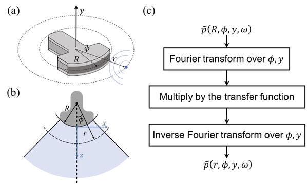
Figure 1. Workflow of the helical wave spectrum (HWS) method for passive acoustic mapping (PAM).
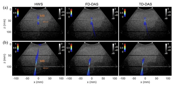
Figure 2. Comparison of the HWS, frequency-domain (FD) and time-domain (TD) delay-and-sum (DAS) methods for PAM.
*This article is provided by Prof. Cai Xiran

 沪公网安备 31011502006855号