
As a novel electromagnetic imaging technique that combined the feature of microwave imaging and ultrasound imaging, microwave-induced thermoacoustic tomography (MITAT) has seen a rapid growth in numerous biomedical applications during the past decades. For the application in transcranial brain hemorrhage detection, the major challenge of MITAT is the big acoustic inhomogeneity induced by the skull. This can cause strong refraction, phase distortion and attenuation in thermoacoustic signals, and therefore, rendering the conventional MITAT algorithms infeasible for transcranial brain hemorrhage detection.
Recently, Associate Professor Wang Xiong’s group in SIST has made significant progress in the research of MITAT. The researchers addressed the adverse effect of the acoustic heterogeneity by using a deep-learning-based MITAT (DL-MITAT) approach for transcranial brain hemorrhage detection. Their achievement was recently published in IEEE Transactions on Biomedical Engineering in an article entitled “Deep-learning-enabled Microwave-induced Thermoacoustic Tomography based on ResAttU-Net for Transcranial Brain Hemorrhage Detection”.
In the study, the researchers proposed a new network structure, a residual attention U-Net (ResAttU-Net), for the DL-MITAT technique, and exhibited an improved performance as compared to some traditionally used networks. By using a 3D-printed skull, an 8.1-mm thick bovine skull and porcine brain tissues to perform ex vivo experiments, the researchers have demonstrated that the trained ResAttU-Net is capable of efficiently eliminating image artifacts and accurately restoring the hemorrhage spot. They also used the CST simulation software to make EM-thermal coupling simulations on a 3D realistic adult human head to study the energy and temperature level for practical scenarios.
In recent years, the huge success achieved by deep learning methods in medical imaging has been widely witnessed. Unlike traditional imaging methods, MITAT as a novel imaging technique does not have a large amount of open-source clinically obtained databases, so it is one of the main challenges in applying deep leaning technique on MITAT. The DL-MITAT technique for hemorrhage detection proposed by Wang’s group utilized simulation methods to establish training sets. This not only fills the technical gap in this field, but also has the potential to be extended to other applications of MITAT and other microwave imaging modalities.
SIST third-year master student Li Chenzhe is the first author of this paper, SIST second-year master student Xi Zijun is thesecond author. Prof. Wang Xiong is the corresponding author.
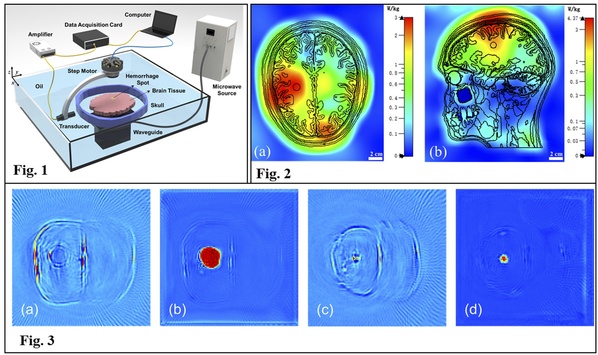
Figure 1.Schematic of experiment system. Figure 2. SAR distribution in the head. (a) transverse plane. (b) sagittal plane. Figure 3. Printed-skull-based ex vivo experimental testing results. (a) and (b) are respectively the DAS and recovered images for the sample with a 10-mm-diameter hemorrhage spot. (c) and (d) are respectively the DAS and recovered images for the sample with a 5-mm-diameter hemorrhage spot.
**This article is provided by Prof. Wang Xiong

 沪公网安备 31011502006855号